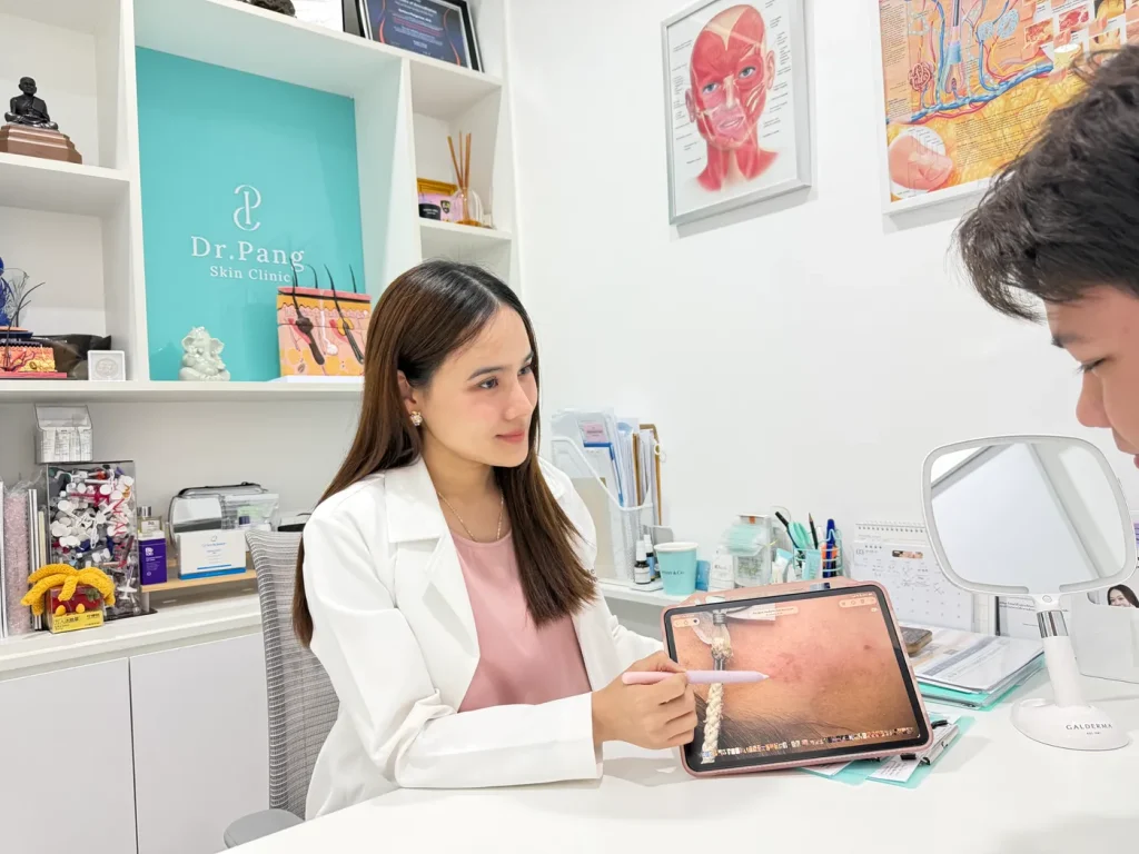
A skin cancer screening in Bangkok takes 15 to 30 minutes, depending on how much of your skin is checked. There are no needles, no cutting, and nothing to prepare. You leave knowing which spots are safe, which need watching, and which need a test. Most people leave with nothing more than a follow-up date in their calendar.
What stops most people from booking is not the price. It is not knowing what happens in the room: how much clothing comes off, whether the doctor will cut something out, or what it means if a mole looks suspicious. This article walks through a skin cancer screening at Dr. Pang Skin Clinic step by step, so nothing about the visit surprises you.
The short version
- Duration: 15 to 30 minutes, depending on the scope: focus spot, focal area, or whole-body check.
- Preparation: none. Removing nail polish and makeup helps.
- Pain: none. Dermoscopy is just a glass lens resting on the skin.
- What is examined: your whole skin surface, plus every pigmented or abnormal red/pink lesion under a dermatoscope — not only the ones that look worrying.
- What you leave with: an explanation the same visit, dermoscopic photos of anything being monitored, and a clear next step.
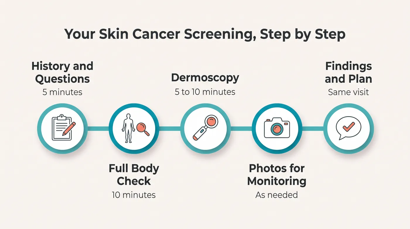
What a skin cancer screening actually is
A screening is a careful head-to-toe examination of your skin by a dermatologist. The goal is to find cancers and pre-cancers while they are still small, shallow, and curable. It is a diagnostic appointment, not a treatment appointment.
That difference matters. A screening does not automatically lead to a procedure. In most visits, nothing is removed at all. If something does need to come out, it is scheduled for later and explained first, because a lesion that might be cancerous must be sampled and sent to a laboratory, never destroyed.
Choosing the scope of your screening
Not every visit needs to cover your whole body. You can choose how much of your skin gets checked:
- Focus spot check: one lesion you are worried about, examined under the dermatoscope.
- Focal area check: a defined area with several lesions, usually three to five, such as a cluster of moles on one arm or across your back.
- Whole-body check: a systematic head-to-toe examination. This is the recommended option if it is your first screening, if you have many moles, or if you cannot remember when each area was last checked.
Not sure which one fits you? Book a whole-body check. It is the only option that also covers the areas you cannot see or reach yourself.
Before your visit: five things that make the appointment better
- Remove nail polish and gel from fingers and toes. Melanoma in Asian skin often appears on the soles, palms, or nail beds, and polish hides the nail bed completely.
- Come without makeup, or be ready to remove it. The face, hairline, and ears are among the highest-risk spots for basal cell and squamous cell carcinoma.
- Untie your hair. The scalp is checked, and it is one of the most commonly missed areas.
- Write down the spots that worry you. People often forget the one lesion they came in about once the examination starts. A photo on your phone is even better.
- Wear clothing that is easy to take off and put back on. You will change into a gown.
Step 1: The conversation before the examination
The first few minutes are about your history, and they shape everything that follows. Your doctor will ask about your personal and family history of skin cancer, how much sun you get, whether you have used tanning beds, whether you take immunosuppressive medication or have had an organ transplant, and whether any lesion has changed recently.
The most useful thing you can say is a sentence that starts with this one has changed. Change over time is the strongest warning sign of skin cancer, and only you have that information.
Step 2: The full-body examination
You change into a gown in private. The examination then moves systematically, usually from the scalp downward, under bright examination lighting. The doctor works through each area and explains what they are doing as they go.
How much clothing comes off
You stay in the gown throughout. Each area is uncovered one at a time and covered again before moving on, so you are never fully exposed. If you are uncomfortable with a particular area being examined, say so and it will be skipped. But know what that means: melanoma can grow on skin that never sees sunlight, so a check that skips areas is only a partial check. A female chaperone is available on request.
The places most people forget
These spots carry more risk than their low profile suggests, and they are the reason a professional check finds things a mirror cannot:
- The scalp, parted section by section
- Behind and inside the ears
- The soles of the feet and the spaces between the toes
- The palms, and the nail beds of both hands and feet
- The back, especially the upper back and shoulder blades
- The back of the neck and the hairline
Step 3: Dermoscopy on every pigmented or abnormal-colour lesion
This is the part of the visit that separates a real screening from a quick glance, and it is where our clinic’s protocol is deliberately stricter than the minimum standard.
A dermatoscope is a handheld device that combines magnification with polarised light. Pressed against the skin, it removes the surface reflection so the doctor can see the pigment pattern, blood vessels, and structure just beneath the surface. It is completely painless: a cool glass plate resting on your skin for a few seconds.
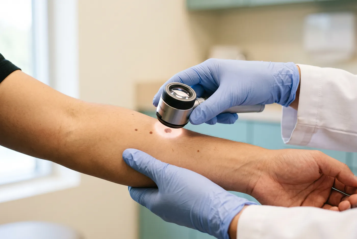
Many clinics pick up the dermatoscope only when a lesion already looks worrying to the naked eye. That approach can only confirm a suspicion you already had, and early melanoma is dangerous precisely because it looks ordinary. At Dr. Pang Skin Clinic, every pigmented lesion and every abnormal red or pink lesion is examined under the dermatoscope, not only the ones that stand out.
Red and pink lesions matter just as much as the brown and black ones. Basal cell carcinoma, the most common skin cancer, usually shows up as a persistent red or pink patch or bump rather than a dark spot, so a check that only looks for pigmented lesions can miss it. Melanoma is rarer than basal cell carcinoma but more aggressive, which is why neither category can be skipped.
I use the dermatoscope on every pigmented or abnormal-colour lesion, not just the frightening ones. The melanomas that get caught early are usually the ones nobody was worried about.
Dr. Gateorn Pongarnar, M.D., Board-Certified Dermatologist and Dermatologic Surgeon
The evidence for this is strong. A meta-analysis of studies done in real clinics found that examination with dermoscopy detected 90% of melanomas, against 71% for the naked eye alone. Dermoscopy also works in the other direction: it correctly identifies benign lesions and prevents unnecessary biopsies.
| Method | Sensitivity for melanoma | What it can see |
|---|---|---|
| Naked eye examination | About 71% | Surface colour, shape, size, and border |
| Examination plus dermoscopy | About 90% | Pigment pattern, blood vessels, and structure beneath the surface |
Dermoscopy also settles most false alarms in seconds. Many of the spots patients arrive worried about turn out to be seborrhoeic keratoses, sun spots, or ordinary benign moles. If the finding is sun-induced pigmentation rather than something dangerous, you will be told so during the same appointment, along with what can be done about it cosmetically if you want.
Step 4: Photographing the lesions worth watching
Not every lesion is clearly benign or clearly malignant. Some are simply uncertain today. For these, your doctor takes a dermoscopic photograph through the instrument and stores it in your record.
That photograph turns an unanswerable question into an answerable one. At your recheck, the new image is compared against the stored baseline. A lesion that looks identical is strongly reassuring. A lesion that has changed gets biopsied. Re-photographing at three months is a well-proven protocol: in published studies, more than 99% of lesions unchanged at three months proved benign on further follow-up, and the method caught 96% of the invasive melanomas among monitored lesions.
This is a quiet advantage of returning to the same clinic. Without a stored baseline image, a doctor seeing you for the first time can only ask whether the mole has changed, and rely on your memory of how it looked a year ago.
Step 5: Findings and plan, explained the same day
You do not wait for a report. Before you leave, your doctor explains what they found and what they recommend. Every screening ends in one of three ways.

| Outcome | What it means | Your next step |
|---|---|---|
| All clear | Nothing looks concerning under dermoscopy | Return in 12 months, sooner if something changes |
| Watch and photograph | One or more lesions are uncertain but not alarming | Dermoscopic image stored today, recheck in 3 months |
| Biopsy recommended | A lesion is suspicious enough that it needs laboratory testing, not more watching | Biopsy scheduled, results in 1 to 2 weeks |
A biopsy recommendation is not a cancer diagnosis. It means the lesion has crossed the line where guessing is no longer acceptable, and only a pathologist examining the tissue can settle it. Most biopsied lesions come back benign.
What happens if a biopsy is recommended
A biopsy is a small procedure under local anaesthetic. You feel the sting of the anaesthetic injection for a few seconds, then pressure but no pain. The technique is chosen to fit the lesion, and the choice matters: the wrong technique can ruin the pathology report.
| Technique | Best suited to | Stitches |
|---|---|---|
| Shave biopsy | Superficial raised lesions | None |
| Punch biopsy | A focused sample to check dermal involvement, usually 3 to 4 mm across and 4 mm deep | 1 to 3 |
| Incisional biopsy | A full-thickness sample of a larger or deeper lesion, which can include the fat layer | Several |
| Excision biopsy | Suspected melanoma, where the whole lesion must come out intact so the margins can be checked | Several |
The sample goes to a dermatopathologist, and results usually return within one to two weeks. If the result confirms cancer, treatment is planned at your results appointment, most often surgical removal with clear margins, confirmed in the laboratory.
One warning belongs here. A lesion that might be cancerous must never be destroyed by laser, cautery, freezing, or a beauty-clinic pen device: destroying it leaves no tissue to examine and no way to know what it was. Benign lesions confirmed by dermoscopy are a different matter: they can be removed in a single session once the diagnosis is settled. The order is what matters: diagnose first, then remove, never the reverse.
How often should you be screened?
| Your risk profile | Screening interval |
|---|---|
| Average risk adult living in Thailand | Every 12 months |
| Fair skin, Fitzpatrick I to II, or more than 50 moles | Every 6 months |
| Personal or family history of skin cancer | Every 6 months |
| Immunosuppressed or organ transplant recipient | Every 3 to 6 months |
| Heavy outdoor sun exposure, or history of blistering sunburn | Every 6 to 12 months |
| A lesion under dermoscopic monitoring | 3 months, then reassess |
Why Bangkok changes the calculation
Bangkok sits at 13 degrees north latitude, and the UV Index regularly reaches 11 to 13 (the extreme band) all year round. There is no low-UV season to recover in. Sun damage builds up faster than people arriving from cooler countries expect, which is why basal cell carcinoma, squamous cell carcinoma, and actinic keratosis are everyday findings in Thai practice.
There is a second, less obvious reason. Melanoma is less common in Asian skin than in fair European skin, and that is exactly why it is dangerous here: lower perceived risk means later diagnosis. When melanoma does occur in Asian and other darker skin, it often takes the acral form, appearing on the palms, soles, and nail beds rather than on sun-exposed skin. Acral lentiginous melanoma is consistently linked to delayed diagnosis and worse outcomes, because public awareness is low and the signs in darker skin are not well recognised.
Those are precisely the spots you cannot easily check yourself, and precisely the spots a full-body screening covers by protocol. Stage matters enormously: five-year relative survival for melanoma caught while localised is over 99%. It falls to about 76% once it reaches nearby lymph nodes, and to roughly 35% once it spreads to distant organs.
What a screening cannot do
Honest limits, because trustworthy information includes them:
- It is not a guarantee for the next twelve months. A melanoma can appear weeks after a clear check. Monthly self-checks are still necessary between visits.
- Dermoscopy is not a diagnosis. It is a very good filter. Laboratory examination of tissue is the only definitive answer, which is why the biopsy threshold exists.
- It does not examine internal organs or mucosal surfaces. Rare mucosal and eye melanomas fall outside a skin check.
- It cannot see areas you decline to have examined. Skipped regions are genuinely unscreened, not presumed clear.
Frequently asked questions
Does a skin cancer screening hurt?
No. A screening is entirely non-invasive. The dermatoscope is a glass lens resting lightly on the skin for a few seconds at a time. Nothing is injected, cut, or frozen. Discomfort only enters the picture if you go on to have a biopsy, and that is done under local anaesthetic at a separate, scheduled appointment.
How long does the appointment take?
Fifteen to thirty minutes, depending on whether you choose a focus spot, focal area, or whole-body check. A whole-body screening runs longer if you have many moles, because every pigmented or abnormal red lesion is checked individually under dermoscopy, or if several lesions need photographing.
Will something be removed on the day?
Usually not. Screening and treatment are separate steps by design: if a lesion is highly suspicious, a biopsy is explained and scheduled for a later date, so you have time to ask questions and plan the wound care. That said, if the schedule allows, a biopsy for a suspected lesion or a CO2 laser removal for a benign, cosmetic lesion can sometimes be arranged on the same day. Ask at booking if this matters to you.
Do I have to take all my clothes off?
You change into a gown, and areas are uncovered one at a time and covered again before moving on. You are never fully exposed. You may decline examination of any area, and a female chaperone is available on request. Bear in mind that any area you skip is genuinely not screened.
I only have one mole I am worried about. Do I need a full-body check?
You can book a focus spot check for just that lesion, and that is far better than doing nothing. But the lesion patients worry about is often benign, while the one that matters is somewhere they have never looked, such as the back, scalp, or sole. A focal area check is a middle option if you would rather cover a defined region than the whole body. Either way, if you are already in the clinic and in a gown, upgrading to a whole-body check takes only a few more minutes.
Is skin cancer screening worth it for Asian skin?
Yes. More natural pigment lowers melanoma risk but does not remove it, and it gives no protection at all on the palms, soles, and nail beds where acral melanoma arises. Basal cell and squamous cell carcinoma are common in Thai patients because of constant extreme UV exposure. The survival gap between early and late detection is wider in groups who present late, which makes screening more valuable here, not less.
What if I am visiting Bangkok as a tourist or expat?
A screening is a single visit and the findings are explained the same day, so it fits easily into a short stay. If a biopsy is needed, the sample takes one to two weeks to report, so allow for that when planning your trip. Bring photographs of any previous mole mapping if you have them.
How should I check my own skin between visits?
Once a month, in good light, using a hand mirror or a partner for your back. Apply the ABCDE rule (asymmetry, irregular border, uneven colour, diameter over 6 mm, and evolution), and also look for the ugly duckling: the one lesion that looks different from all your others. Any change in size, shape, or colour, or new bleeding, itching, or crusting, deserves an appointment without waiting for your annual check.
Booking a screening in Bangkok
Screenings at Dr. Pang Skin Clinic are performed by our team of board-certified dermatologists, following the screening protocol developed by Dr. Gateorn Pongarnar, M.D., a board-certified dermatologist and dermatologic surgeon with more than 13 years of clinical experience. The clinic is at Bangkok Mediplex in Ekkamai, a short walk from BTS Ekkamai.
You can review the clinic’s full skin cancer diagnosis and treatment services, or book a screening appointment directly.
Medical review
This article was written and clinically reviewed by Dr. Gateorn Pongarnar, M.D., Board-Certified Dermatologist and Dermatologic Surgeon at Dr. Pang Skin Clinic, Bangkok. It describes the screening protocol used at this clinic and is intended as general education, not as a substitute for an individual medical assessment. If you have a lesion that is changing, bleeding, or new, seek evaluation promptly rather than waiting for a scheduled screening.
References
- Vestergaard ME, Macaskill P, Holt PE, Menzies SW. Dermoscopy compared with naked eye examination for the diagnosis of primary melanoma: a meta-analysis of studies performed in a clinical setting. British Journal of Dermatology.
- Dinnes J, Deeks JJ, Chuchu N, et al. Dermoscopy, with and without visual inspection, for diagnosing melanoma in adults. Cochrane Database of Systematic Reviews.
- Menzies SW, Gutenev A, Avramidis M, et al. Assessment of the optimal interval for and sensitivity of short-term sequential digital dermoscopy monitoring for the diagnosis of melanoma. Archives of Dermatology.
- Deinlein T, Michor C, Hofmann-Wellenhof R, Schmid-Zalaudek K, Fink-Puches R. The importance of total-body photography and sequential digital dermatoscopy for monitoring patients at increased melanoma risk. Journal der Deutschen Dermatologischen Gesellschaft, 2020.
- American Cancer Society. Survival rates for melanoma skin cancer, by stage (SEER data).
- Trends in skin cancer incidence in Songkhla, Southern Thailand, 1989 to 2020: a population-based study on the impact of geographic variation. PLOS One.
- Acral melanoma in skin of color: current insights and future directions, a narrative review. Cancers.
- Acral lentiginous melanoma: overcoming diagnostic delays through early detection strategies. Journal of the American Academy of Dermatology.
- American Academy of Dermatology. The naked truth about total body skin examination. Dermatology World Insights and Inquiries.